Ultra-High Field Magnetic Resonance Imaging: Towards an Imaging Based Healthcare
We would like to warmly thank Prof. Alayar Kangarlu, Director of the physics and engineering group at NYSPI MRI research center at Columbia University, for his visit to the ISTP and his interesting talk on ultra-high field magnetic resonance imaging and their importance in medicinal research and diagnostics.
by Gian Luca Gehwolf

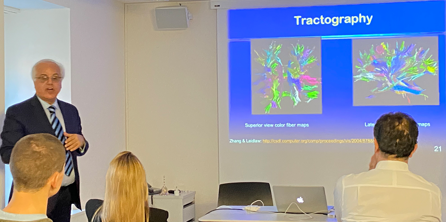
Prof. Kangarlu started his talk on ultra-high field magnetic resonance imaging (MRI) by showing the periodic table, and highlighting the importance of the element Hydrogen as the reason, which made MRI possible in the first place. “The element Hydrogen is a gift from nature”, he stated. The human body mostly comprises of water, which consists of the elements Hydrogen and Oxygen. Additionally, Hydrogen atoms do not have neutrons in their nucleus (they only consist of one proton and one electron). These two reasons enabled obtaining images of the human tissue by using MRI.
The use of magnetic resonance imaging has become an indispensable tool in medicine as it provides high-resolution images of anatomy, physiology and biochemistry of the human body. Unlike cameras for photography, ultra-high magnetic field MRI allows taking different types of images. By fine-tuning the magnetic field, certain classes of tissues can be ignored, e.g. grey matter, white matter, blood, etc., and only the tissue of interest can remain visible.
In the discussion, which followed his talk, the difference of the US and the European healthcare system, regarding the use of MRI, was analysed. Prof. Kangarlu explained that the American system is much more entrepreneurship-orientated compared to the European, which has a more socialist approach with lower costs for the patients to bear.
The ISTP would like to thank Prof. Kangarlu for giving such an interesting talk about the importance of ultra-high field MRI in the healthcare system.
For more information about the talk and the full report, please visit our Reports page.